
Bruker TruLive3D Imager Light-Sheet Microscope
(I) Description
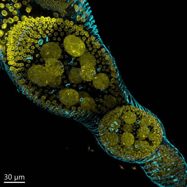
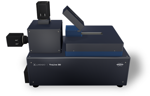
The Bruker Luxendo TruLive3D Imager is an inverted light-sheet fluorescence microscope optimized for fast, long-term 3D imaging of delicate live specimens in their native biological environment. Its dual-sided illumination and single-lens detection from below enable rapid acquisition, deep imaging, and uniform illumination with minimal shadowing, while advanced environmental control helps preserve sample viability during experiments lasting from hours to days. A large multiplex sample holder supports hundreds of organoids, spheroids, 3D cell cultures, or small embryos and enables comparison of up to six experimental conditions within a single experiment.
(II) Features & Benefits
- Fast, low-phototoxicity 3D imaging of delicate live specimens
- Dual-sided illumination for deep, uniform imaging with reduced shadowing
- High-NA detection for excellent sensitivity and spatial resolution
- Long-term imaging under controlled physiological conditions
- Multiplex acquisition of hundreds of samples and up to six experimental conditions
- Dual-destriping and TAG-lens (tunable acoustic gradient lens) technologies for improved light-sheet uniformity
- Easy open-top sample access and flexible TruLive3D sample holders
- Optional photomanipulation for advanced functional imaging experiments
- Integrated LuxBundle software for acquisition, processing, and 3D visualization
These capabilities are designed particularly for multi-sample and multi-condition live-imaging workflows involving organoids, spheroids, cell cultures, and small embryos.
(III) Technical Specifications
- Imaging modality: Inverted Light-Sheet Fluorescence Microscopy with dual-sided illumination and single-lens detection
- Light-sheet generation: Beam-scanned Gaussian light sheets
- Light-sheet thickness: Adjustable from 2 to 6 µm
- Illumination optics: Chromatically corrected from 440 to 660 nm
- Illumination objectives: 2 × Nikon CFI Plan Fluor 10× W, 0.3 NA water-immersion objectives
- Detection objective: Nikon CFI Apo 25× W, 1.1 NA water-immersion objective
- Alternative objective configuration: Olympus XLUMPLFLN 16×, 0.8 NA
- Effective magnification: Multiple configuration-dependent options from approximately 8× to 62.5×
- Detection: 2 × Hamamatsu ORCA-Flash4.0 V3 high-speed sCMOS cameras
- Camera resolution: 2048 × 2048 pixels with 6.5 × 6.5 µm pixel size
- Acquisition speed: More than 80 frames per second at full frame and up to 500 frames per second with subframe cropping
- Peak quantum efficiency: Up to 82% at 560 nm
- Spectral detection: Two detection channels with 10-position high-speed emission filter wheels
- Simultaneous imaging: Three motorized dichroic positions for simultaneous dual-channel acquisition
- Illumination control: Motorized neutral-density filters with 90%, 10%, and 1% transmission
- Environmental control: Temperature from 20 to 37 °C, CO₂, O₂/N₂, and humidity control
- Sample capacity: 75 mm sample holder accommodating up to hundreds of specimens
- Experimental conditions: Up to six separate conditions using three dual-compartment TruLive3D dishes
- Sample mounting: FEP-foil-based sample holders for cells, organoids, spheroids, and embryos
- Stage travel: 66 × 3 × 3 mm
- Artifact reduction: Dual-destriping through pivot scanning
- Light-sheet optimization: TAG-lens technology for rapid axial scanning and illumination uniformity
- Photomanipulation: Optional photomanipulation module
- Software: Bruker LuxBundle for acquisition, multidimensional experiment control, post-processing, and 3D visualization
- System design: Compact, vibration-free benchtop configuration without the need for an air table
Bruker’s published objective, magnification, and field-of-view values depend on the selected optical configuration; the final configuration should therefore be confirmed when preparing a quotation or formal datasheet.
(IV) Manufacturer: Bruker
(V) Applications
Live-Cell Imaging, Long-Term Time-Lapse Imaging, 3D Cell Culture Imaging, Organoid Imaging, Spheroid Imaging, High-Throughput Multi-Sample Imaging, Multi-Condition Drug Studies, Developmental Biology, Embryo Development, In Vivo Imaging of Small Model Organisms, Zebrafish Research, Drosophila Research, Mouse Embryo Imaging, Cancer Biology, Tumorigenesis, Drug-Response Studies, Toxicology, Stem Cell Research, Primary Cell Imaging, Cell–Cell Interaction Studies, Immunology, Wound-Healing Studies, Photomanipulation.
Related products