

BC43 – Ultimate Benchtop Confocal Microscope | Andor
Spinning disk confocal microscopy in a compact, automated and accessible system
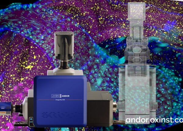
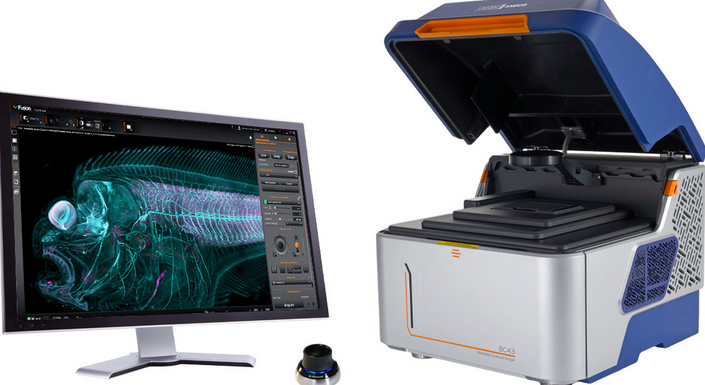
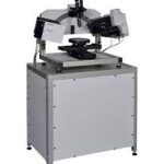
Andor BC43 is a compact microscopy platform for widefield fluorescence, spinning disk confocal, transmitted-light and super-resolution imaging. It is designed for laboratories requiring high-quality 2D and 3D imaging without the operational complexity and physical footprint of a conventional confocal microscope.
Its fully enclosed, light-tight design allows BC43 to operate on a standard laboratory bench without a dedicated dark room. An integrated vibration-reduction mechanism and automated optical components support consistent results, including for researchers with limited previous experience in advanced microscopy.
BC43 can image fixed and live cells, 3D cell cultures, organoids, embryos, model organisms, tissue sections and large biological specimens. Confocal configurations use spinning disk technology derived from Andor’s Dragonfly platform to provide rapid acquisition and effective optical sectioning.
The platform is available in several configurations:
- BC43 WF for widefield fluorescence and transmitted-light imaging;
- BC43 CF for widefield, spinning disk confocal and transmitted-light imaging;
- BC43 SR with optional super-resolution capability;
- field upgrades from widefield to confocal and from standard imaging to super-resolution.
APEL Laser can assist with the selection of the BC43 configuration, objectives, imaging modalities, software modules and analysis tools required for your research.
Benefits of the Andor BC43
Fast confocal imaging
BC43 CF uses a spinning disk confocal architecture with microlens and pinhole arrays. Multiple points within the specimen are illuminated and detected simultaneously, rather than scanning one point sequentially across the sample.
This enables:
- rapid confocal image acquisition;
- efficient Z-stack imaging;
- visualisation of dynamic biological processes;
- shorter total exposure;
- increased throughput for large samples and volumes;
- gentler imaging of living cells and model organisms.
Andor reports productivity of at least ten times that of certain point-scanning confocal systems, depending on the experiment and system configuration.
Gentle live-cell imaging
Parallel illumination, efficient light collection and a sensitive sCMOS camera reduce the exposure required for each acquisition.
BC43 can support long-term observation of:
- cell division;
- migration;
- embryonic development;
- intracellular transport;
- organelle dynamics;
- cellular signalling;
- cell-to-cell interactions;
- processes within model organisms.
The system is designed to image live specimens over extended periods while reducing photobleaching and illumination-induced disturbance.
Fully enclosed system
The light-tight enclosure allows the microscope to operate without a dark room.
This provides:
- isolation from ambient light;
- consistent acquisition conditions;
- protection of the specimen and optics;
- reduced contamination;
- simplified use in shared laboratories;
- a compact installation footprint.
Accessible operation
BC43 is intended for users with different levels of microscopy experience. Automated focus, objective selection, illumination, filtering, stage movement and image acquisition minimise manual adjustment.
Andor indicates that a new user can typically become independent within approximately 30 minutes to two hours, depending on the complexity of the experiment and the training provided.
BC43 configurations
BC43 WF – upgradeable widefield microscopy
BC43 WF is intended for laboratories performing predominantly fluorescence imaging of thin specimens or low-signal samples.
The configuration can include:
- laser-based widefield fluorescence;
- brightfield;
- Differential Phase Contrast;
- ClearView™ GPU deconvolution;
- multichannel acquisition;
- Z-stacks;
- time-lapse;
- montage and multipoint imaging;
- analysis in Imaris.
BC43 WF can later be upgraded in the field to the confocal model without replacement of the complete microscope.
BC43 CF – spinning disk confocal microscopy
BC43 CF adds spinning disk confocal imaging for optical sectioning and three-dimensional imaging of thicker specimens.
It is recommended for:
- organoids;
- spheroids;
- embryos;
- tissue sections;
- 3D models;
- neuroscience;
- developmental biology;
- model-organism imaging;
- specimens requiring effective rejection of out-of-focus fluorescence.
BC43 CF retains widefield and transmitted-light modalities, enabling the appropriate method to be selected for each experiment.
BC43 SR – accessible super-resolution
BC43 SR uses Andor’s optimised SRRF-Stream+ technology to produce resolution beyond the conventional diffraction limit.
The manufacturer specifies:
- resolution down to approximately 140 nm;
- an improvement of approximately 1.5–1.7 times over conventional imaging;
- compatibility with standard labelling protocols;
- use with thick or thin samples;
- compatibility with widefield and confocal modalities;
- suitability for live-cell protocols.
Super-resolution imaging can reveal actin filaments, microtubule networks, vesicles, lysosomes and protein interactions more clearly.
Configuration comparison
| Feature | BC43 WF | BC43 CF | BC43 SR |
|---|---|---|---|
| Widefield fluorescence | Yes | Yes | Yes |
| Spinning disk confocal | Field upgrade | Yes | Depends on base configuration |
| Brightfield | Yes | Yes | Yes |
| Differential Phase Contrast | Yes | Yes | Yes |
| ClearView GPU deconvolution | Available | Available | Available |
| 2D and 3D imaging | Yes | Yes | Yes |
| Thick specimens | Limited | Yes | Yes |
| Super-resolution down to approximately 140 nm | Upgrade | Upgrade | Yes |
| Field upgradable | Yes | Yes | Yes |
| Primary use | Thin specimens and routine imaging | 3D specimens and confocal imaging | Fine subcellular structures |
The exact capability should be confirmed according to the selected model, installed modules and software licences.
Imaging modalities
Spinning disk confocal microscopy
Confocal imaging rejects much of the out-of-focus fluorescence and optically separates different planes within a specimen.
It is suited to:
- 3D imaging;
- multicellular samples;
- three-dimensional cultures;
- tissues;
- embryos;
- model organisms;
- depth-resolved protein localisation.
Widefield fluorescence
Widefield provides high sensitivity and maximum speed for relatively thin specimens.
It can be used for:
- 2D cell cultures;
- weak fluorescence signals;
- rapid biological processes;
- screening;
- time-lapse imaging;
- live-cell monitoring;
- acquisitions not requiring confocal optical sectioning.
Brightfield
Brightfield provides label-free visualisation of specimens with natural contrast.
It is useful for:
- model organisms;
- general specimen morphology;
- sample positioning and orientation;
- tissue-boundary visualisation;
- correlation of structural and fluorescence data.
Differential Phase Contrast
DPC improves contrast in transparent or weakly contrasted specimens.
It is suitable for:
- unlabelled cells;
- transparent samples;
- culture inspection;
- morphology monitoring;
- correlation of structural and fluorescence information.
SRRF-Stream+ super-resolution
SRRF-Stream+ combines information from multiple image frames and analyses local intensity fluctuations to reconstruct features that cannot be separated by conventional imaging.
Compared with single-molecule localisation methods, it can operate with labelling procedures that are closer to routine fluorescence protocols and with a simpler experimental workflow.
Excitation sources and imaging channels
BC43 uses four laser excitation lines:
- 405 nm;
- 488 nm;
- 561 nm;
- 640 nm.
These wavelengths cover many commonly used fluorophores, including nuclear stains, green and red fluorescent proteins and far-red fluorescent dyes.
The system can provide up to ten imaging channels:
- four widefield channels;
- four confocal channels;
- two transmitted-light modalities.
Fluorophore selection should consider excitation wavelengths, emission filters and the spectral separation required between channels.
Objectives and focusing
BC43 can be configured with up to five interchangeable objectives.
Objective selection should consider:
- specimen dimensions;
- required field of view;
- target resolution;
- specimen thickness;
- working distance;
- immersion medium;
- signal brightness;
- slide, plate or vessel format.
The system can accommodate objectives for large fields and whole organisms as well as high-numerical-aperture objectives for subcellular structures.
The motorised XYZ stage supports:
- multipoint acquisition;
- Z-stacks;
- tiled imaging;
- automated specimen navigation;
- repeated imaging at predefined positions;
- multiwell-plate workflows.
Large-sample imaging
BC43 can combine multiple image fields and volumes to visualise specimens larger than the field of a single objective.
Fusion Benchtop capabilities include:
- XY montage;
- XYZ stitching;
- spiral montage;
- irregular montage;
- Focus Map;
- Z-intensity correction;
- multipoint acquisition;
- 3D time-lapse imaging.
Irregular Montage acquires only relevant regions, reducing acquisition time and data volume.
Focus Map compensates for specimen tilt and variations in sample height, keeping selected areas within the focal range.
Applications
Cell biology
BC43 can support studies involving:
- cell division;
- cytoskeletal organisation;
- mitochondria;
- vesicles and lysosomes;
- intracellular transport;
- migration;
- adhesion;
- autophagy;
- protein localisation;
- live-cell imaging.
Developmental biology
Its imaging speed and capacity to acquire large volumes make BC43 suitable for:
- embryos;
- Drosophila;
- zebrafish;
- organ development;
- morphogenesis;
- early cell division;
- cell-lineage analysis;
- 3D time-lapse experiments.
Neuroscience
Applications can include:
- neurons and glial cells;
- neuronal processes;
- dendritic spines;
- synapses;
- axonal transport;
- neuronal networks;
- brain sections;
- three-dimensional neural samples.
Available Imaris modules can support filament tracing, spine identification and quantitative neuronal morphology.
Cancer research
BC43 can be used to investigate:
- proliferation;
- genome instability;
- migration and invasion;
- treatment response;
- 3D tumour cultures;
- organoids;
- biomarker distribution;
- cellular and nuclear morphology.
Organoids and 3D cultures
Confocal imaging optically separates planes within samples that are thicker than conventional monolayer cell cultures.
BC43 can be applied to:
- intestinal organoids;
- cerebral organoids;
- tumour models;
- spheroids;
- hydrogel cultures;
- tissue models;
- volumetric and morphological analysis.
Immunology and microbiology
The platform can support:
- cell-to-cell interactions;
- immunological synapses;
- phagocytosis;
- microorganism localisation;
- biofilms;
- inflammatory responses;
- cell-population analysis;
- multichannel colocalisation.
Core imaging facilities
Simplified operation and the optional IQ/OQ programme make BC43 suitable for shared facilities used by researchers with different levels of microscopy expertise.
Benefits include:
- rapid user training;
- standardised protocols;
- reproducibility between users;
- efficient instrument utilisation;
- documented system performance;
- future upgrades.
Fusion Benchtop software
Fusion Benchtop controls the microscope and enables multidimensional acquisition protocols.
Functions can include:
- imaging-mode selection;
- laser and channel control;
- objective selection;
- XYZ-stage control;
- autofocus;
- multichannel imaging;
- Z-stacks;
- time-lapse;
- multipoint acquisition;
- montage;
- 3D stitching;
- protocol-based deconvolution;
- real-time 3D visualisation;
- Focus Map;
- Irregular Montage;
- depth-intensity correction.
ClearView™ deconvolution can be activated directly within the acquisition protocol, reducing the need for separate manual post-processing.
Image analysis with Imaris
Andor Benchtop microscopes include the Imaris Quant package for image visualisation and analysis.
Imaris can be used for:
- 2D, 3D and 4D visualisation;
- object segmentation;
- intensity-based or AI-assisted object identification;
- object counting;
- volume and morphology measurements;
- fluorescence-intensity quantification;
- creation of images and animations;
- comparison of experiments;
- analysis of biological surfaces and structures.
Additional modules are available for tracking, motility analysis, cell and organelle quantification and neuronal tracing.
IQ/OQ quality and reproducibility
Andor offers an optional Installation Qualification and Operational Qualification programme for BC43.
The programme can assess:
- laser power;
- illumination uniformity;
- illumination centring;
- channel co-registration;
- detector response;
- 3D reconstruction accuracy;
- XY-stage repeatability;
- lateral and axial resolution;
- system vibration;
- optical contamination and background.
For BC43 systems evaluated with a 60×, 1.42 NA objective, Andor publishes OQ specifications of no more than 280 nm average confocal lateral resolution and no more than 725 nm average axial resolution.
Periodic testing provides quantitative information on instrument performance and can support the reproducibility of experiments between users, dates and locations.
Why choose Andor BC43?
BC43 is suitable for laboratories seeking the capabilities of an automated imaging system in an accessible, compact platform.
Its main benefits include:
- fast spinning disk confocal imaging;
- widefield and transmitted light in one instrument;
- optional super-resolution;
- 2D, 3D and 4D imaging;
- fixed- and live-specimen imaging;
- reduced phototoxicity;
- fully enclosed design;
- no dark room required;
- integrated vibration reduction;
- four laser lines;
- up to five objectives;
- motorised XYZ stage;
- multipoint and montage acquisition;
- GPU deconvolution;
- Fusion Benchtop software;
- Imaris analysis;
- optional IQ/OQ programme;
- field upgrades from widefield to confocal and super-resolution.
Configuration and support from APEL Laser
APEL Laser can help define a BC43 configuration tailored to your application.
Configuration support can include:
- selection of BC43 WF, CF or SR;
- definition of imaging modalities;
- objective selection;
- fluorophore assessment;
- field-of-view and resolution requirements;
- live-cell configuration;
- selection of slide, plate or vessel holders;
- multipoint workflow definition;
- selection of Fusion modules;
- selection of Imaris packages;
- installation and calibration;
- user training;
- protocol development;
Frequently asked questions
What is Andor BC43?
BC43 is a compact, automated fluorescence microscope available in widefield, spinning disk confocal and super-resolution configurations.
What is the difference between BC43 WF and BC43 CF?
BC43 WF is intended for widefield imaging of thin and light-sensitive specimens. BC43 CF adds spinning disk confocal imaging for optical sectioning and imaging of thicker samples.
Can BC43 WF be upgraded to confocal?
Yes. Andor provides an in-field upgrade path from BC43 WF to BC43 CF.
Does BC43 provide super-resolution?
Yes. The SRRF-Stream+ module can achieve resolution down to approximately 140 nm, depending on the specimen, fluorophore, objective and acquisition conditions.
Is it suitable for live-cell imaging?
Yes. Rapid acquisition and efficient light collection support live imaging with reduced illumination dose.
Can it image organoids and tissues?
BC43 CF is suitable for 3D specimens, organoids, embryos and tissue sections. Accessible imaging depth depends on the specimen, labelling, objective and tissue optical properties.
Is a dark room required?
No. BC43 has a light-tight enclosure and can operate on a standard laboratory bench.
Which laser wavelengths are included?
The system uses four excitation wavelengths: 405, 488, 561 and 640 nm.
Can it perform multipoint and montage imaging?
Yes. Its motorised XYZ stage and Fusion software support multipoint, montage, stitching, Focus Map and multidimensional acquisition.
Is analysis software included?
Andor Benchtop microscopes include Imaris Quant, with additional modules available according to the application.
Related products